Ultrafine Ferritin-based Iron Oxide Nanoparticles Used for MRAThe high spatial resolution, excellent anatomic details and noninvasive diagnostic process of magnetic resonance imaging (MRI) make it a pivotal technology in clinical imaging. One of the important applications is its blood pool imaging, termed magnetic resonance angiography (MRA), and in particular the use of safe and effective MRI contrast agents to enhance MRA has attracted much attention. Prof. Pan Yongxin's laboratory observed that the positive MRA can be enhanced by using ultrafine ferritin-based iron oxide nanoparticles. This work was published in Nanoscale and was selected as a cover article. The main function of MRI contrast agents is to shorten the protons' longitudinal relaxation time (T1), thereby resulting in brighter images, or to decrease the protons' transverse relaxation time (T2) to generate darker images. The two are usually defined as T1 (positive) or T2 (negative) contrast agents, respectively. The mainstream MRI T1 contrast agents used in clinics are Gd chelates. However, the Gd-based contrast agents have certain limitations, including the risk of adverse effects such as nephrogenic systemic fibrosis, deposition in the brain, and short circulating time. Iron oxide nanoparticles show good biocompatibility, and are typically exploited as T2 contrast agents due to their large transversal relaxivity (r2). Recently, ferritin-based magnetoferritin (M-HFn) nanoparticles were used as T2 contrast agents, and were found to be very promising reagents in tumor detection and imaging. Can such nanoparticles be used to develop a Gd-free T1 contrast agent? Under the guidance of Prof. Pan Yongxin, Cai Yao and other members of the laboratory synthesized ultrafine ferritin-based iron oxide nanoparticles with hematite and/or maghemite cores (diameters < 5 nm), through strictly controlling iron loading into the genetic expressed human H chain ferritin. Among them, the M-HFn-2.2 nanoparticles have a high r1 value of 0.86 mM-1 s-1. In the MRA experiments, high spatial resolution steady-state images were acquired in the vascular nets from 3 minutes up to 2 hours after a single injection of the M-HFn-2.2 nanoparticles, mainly thanks to long-term blood pool imaging. It has been also found that those injected M-HFn-2.2 nanoparticles were mainly distributed in the liver, spleen and kidney within one day post-injection, but no abnormal change was observed in the histopathological examination. The single injection and long-term imaging features suggest their obvious advantages in MRA application compared with other contrasts. Due to their capacities of precisely finely tuned grain size, biocompatibility and active targeting, ultrafine M-HFn nanoparticles are promising in developing clinically applicable Gd-free T1 contrast agents. This study is also a new development of ferritin-based multifunctional nanomaterials to be used for theranostics. This work is supported by the National Natural Science Foundation of China, Key Program of Chinese Academy of Sciences and China Postdoctoral Science Foundation Funded Project.
Figure 1: MRA can be enhanced up to 2 hours by single injection of ferritin-based nanoparticles with hematite/maghemite inner cores. (Image by Prof. PAN Yongxin's group)
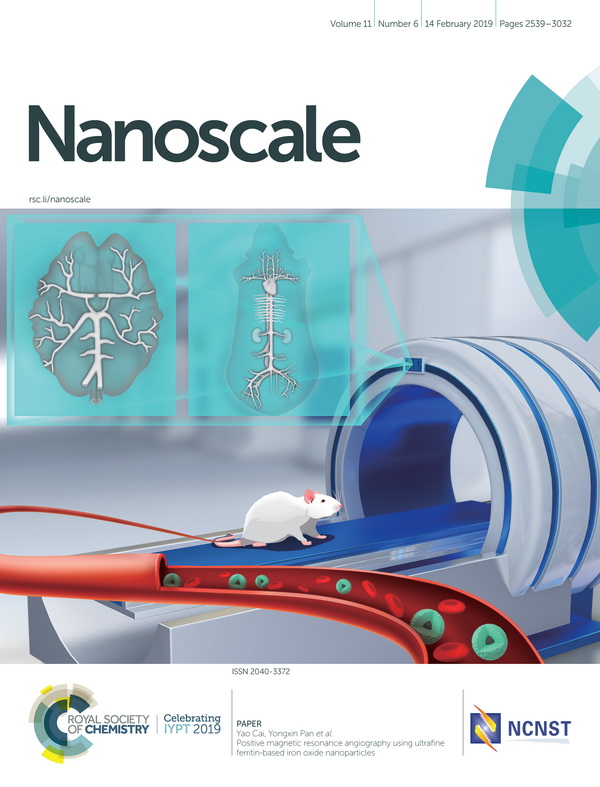
Figure 2: Front cover of Nanoscale, 2019, Issue 6.
|
Contact
CAI Yao
The Institute of Geology and Geophysics, Chinese Academy of Sciences E-mail: caiyao@mail.iggcas.ac.cn PAN Yongxin Phone: +86-010-82998406 The Institute of Geology and Geophysics, Chinese Academy of Sciences E-mail: yxpan@mail.iggcas.ac.cn Related Articles
Reference
|
 Print
Print Close
Close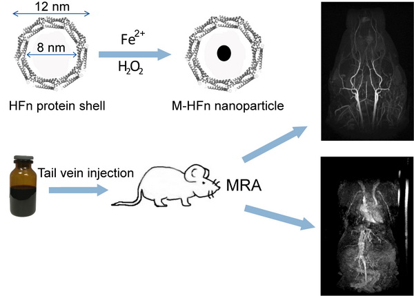
-
SIMSSecondary Ion Mass Spectrometer Laboratory
-
MC-ICPMSMultiple-collector ICPMS Laboratory
-
EM & TEMElectron Microprobe and Transmission Electron Microscope Laboratory
-
SISolid Isotope Laboratory
-
StIStable Isotope Laboratory
-
RMPARock-Mineral Preparation and Analysis
-
AAH40Ar/39Ar & (U-Th)/He Laboratory
-
EMLElectron Microscopy Laboratory
-
USCLUranium Series Chronology Laboratory
-
SASeismic Array Laboratory
-
SEELaboratory of Space Environment Exploration Laboratory
-
PGPaleomagnetism and Geochronology Laboratory
-
BioMNSFrance-China Bio-mineralization and Nano-structure Laboratory