Research and applications of magnetic nanoparticles are very important in rock magnetism, environmental magnetism, biomedicine and nano-materials. Biogenic magnetic nanoparticles, in particular, are important due to their high crystallinity, uniform particle size and good biocompatibility. Ferritin is an iron storage protein that is ubiquitously distributed throughout the microbial, plant, and animal kingdoms. It is composed of a spherical cage-like protein shell with an outer (inner) diameter of 12–13 nm (8 nm), which can be effectively used to synthesize ferrimagnetic H-ferritin nanoparticle (M-HFn).
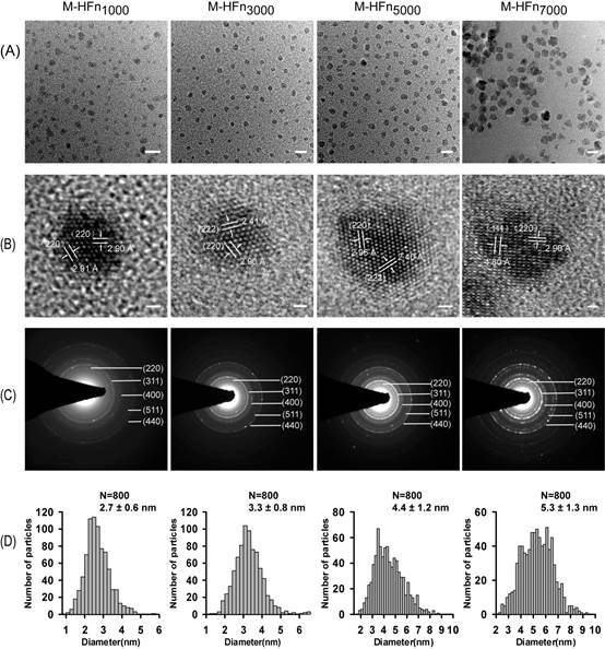
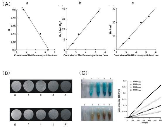
Nano-scale size effects of magnetic nano-particles are important. Recently, doctoral candidate Mr. Yao Cai and his supervisor Prof. Yongxin Pan from the Franco-China Bio-mineralization and Nano-Structures Laboratory, Institute of Geology and Geophysics, CAS systematically investigated the nano-scale size effects of M-HFn. M-HFn nanoparticles with different sizes of magnetite cores in the range of 2.7–5.3nm were synthesized through loading different amounts of iron into recombinant human H chain ferritin (HFn) shells (Figure 1). The study utilized a combination of transmission electron microscope, low-temperature magnetic measurements, magnetic resonance imaging of tumor cells and immunohistochemical staining of tumor tissues. Through these methods, they found that the saturation magnetization (Ms), relaxivity, and peroxidase-like activity of synthesized M-HFn nanoparticles monotonically increased with the size of ferrimagnetic cores (Figure2 A-C). The M-HFn nanoparticles with the largest core size of 5.3 nm exhibit the strongest saturation magnetization, the highest peroxidase activity in immunohistochemical staining, and the highest r2 of 321 mM-1 s-1. This allows the detection of MDA-MB-231 breast cancer cells with concentrations as low as 104 cells mL-1.
M-HFn nanoparticles synthesized here are coated with ferritin shell, monodisperse, contain no obvious lattice defects, are uniform in size and nearly spherical shape; they are ideal model for studying size effects of magnetite at the nano-scale. The group had showed previously that M-HFn can be targeted to tumor cells without modification of the outer ferritin shells; it can be used as a new type of magnetic resonance imaging contrast agent for MRI of tumors. The M-HFn nanoparticles can transport from the blood stream to the target site, while penetrating various biological barriers, which make them sufficiently sensitive to detect tumors with low TNRs (i.e., microscopic carcinomas, <1–2 mm in diameter prior to angiogenesis). The present study demonstrated that, by simply controlling the size of magnetite core, the contrast ability and immunohistochemical staining efficacy of M-HFn nanoparticles can be finely tuned. It may significantly improve the development of new iron oxide contrast agents and rapid diagnostic reagents of tumor tissues.
This work was supported by the CAS/SAFEA International Partnership Program for Creative Research Teams, the National Natural Science Foundation of China, and the R&D of Key Instruments and Technologies for Deep Resources Prospecting. The study was recently published in the International Journal of Nanomedicine (Yao Cai et al. Enhanced magnetic resonance imaging and staining of cancer cells using ferrimagnetic H-ferritin nanoparticles with increasing core size. Int J Nanomed, 2015:10 2619-2634).

Figure 1: TEM analysis of M-HFn nanoparticles. (A) TEM graphs of M-HFn1000, M-HFn3000, M-HFn5000, and M-HFn7000. scale bar is 10 nm. (B) The high-resolution TEM images of these four M-HFn samples. Scale bar is 1 nm. (C) Corresponding SAED patterns of M-HFn nanoparticles. (D) Size histograms of M-HFn nanoparticles.

Figure 2: (A) The relationship between the core size of M-HFn nanoparticles and their magnetostatic interaction parameter (R-value), saturation magnetization (Ms), and coercivity (Hc). (B) MRI of MDA-MB-231 tumor cells incubated with M-HFn nanoparticles. (g)-(k) T2-weighted MR images of tumor cells of different concentrations incubated with M-HFn7000 nanoparticles (5.3 nm). (C) M-HFn nano-particles with different sizes of core catalyzed the oxidation of peroxidase substrates in the presence of H2O2. TMB as the substrate to give a deep blue color product; DAB as the substrate to give a deep brown color product. (a)-(e) in (B) and (C) are control, M-HFn1000, M-HFn3000, M-HFn5000, and M-HFn7000.

 Print
Print Close
Close

